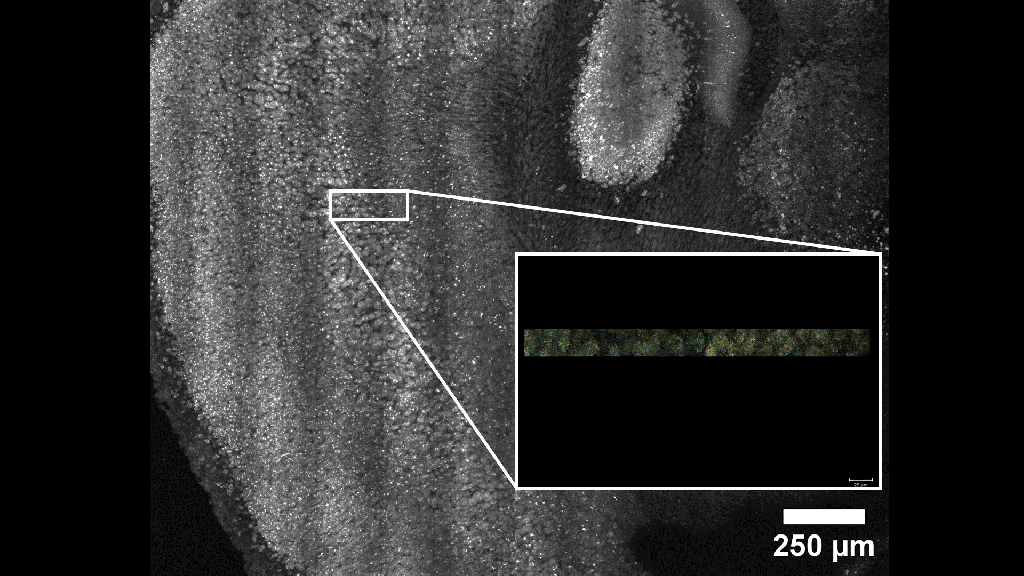
3D multiplexed error resistant RNA fluorescence in situ hybridization (MERFISH)
Maximum projection image of fluorescently labeled cell bodies in 100 um thick mouse brain slice. Inset shows animation of 3D fluorescent image two channels (cyan, orange) of barcoded RNA-FISH throughout the 100 um slice, beginning with XY and transitioning to XZ. Imaged using stage-scanning high-resolution oblique plane microscopy combined with a fluidics controller and depth-dependent spherical aberration compensation.
3D multiplexed immunofluorescence
3D multiplexed immunofluorescence of cleared pediatric brain biopsy. Nuclei (magenta), ILR6a (cyan), beta-catenin (orange), and EPCAM (white). Imaged using stage-scanning high-resolution oblique plane microscopy.
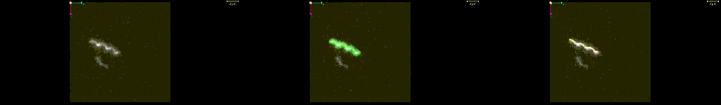
Hydrodynamics of bacterial motility
Propulsion efficiency of individual microscale propellers
3D fluorescence timelapse of Raw (left), segmented (middle), and model quantified (right) isolated, fluorescent labeled, E. coli flagella diffusing in 70:30 water:glyercol mixture. Imaged at 15 volumes/second for 10 seconds using high-resolution oblique plane microscopy.
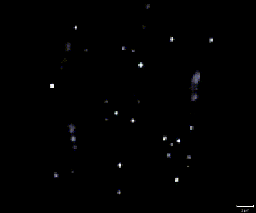
Low Reynolds number swimming dynamics of interacting bacteria
3D refractive index timelapse of E. coli swimming (large objects) and tracer particles diffusing (small objects) in water. Imaged at 150 volumes/second using Fourier-synthesis optical diffraction tomography.
Signal-activated gene and protein expression
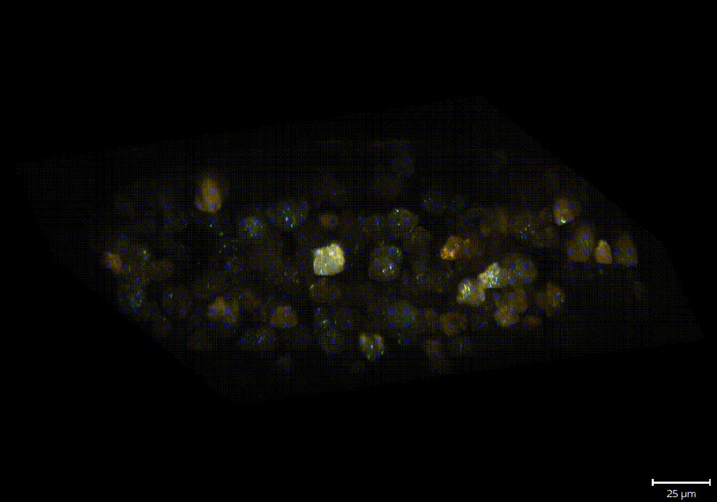
Autophagic flux in 3D cancer cell cultures
3D fluorescence timelapse of matrix-embedded MOLM13 cells expressing GFP(cyan)-mCherry(orange)-LC3. Imaged at 2 volumes/minute for 1 hour using high-resolution oblique plane microscopy.